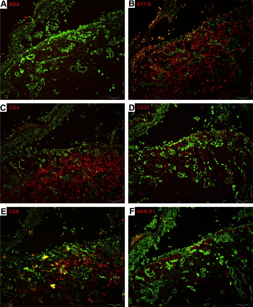
Figure 2. Leukocyte infiltration in GVHD skin.
Antibody tissue staining of a skin lesion from a rat transplanted with 30 × 106 PVG.7B BM cells, which developed lethal aGVHD. Epithelial cell-specific marker isolectin (green color) was added to visualize skin morphology. Negative control staining was done with BSA solution and no primary antibody (A). Skin showed typical signs of grade III-IV GVHD with extensive disruption of the dermal-epidermal interface. Substantial infiltration of donor-derived leukocytes (RT7.B; red color) was noted in the dermis (B). The majority of infiltrating cells expressed CD4 and in fewer numbers CD8, whereas NKR-P1+ cells were virtually absent (C, E, and F). CD25 expression was detected on a minor fraction of leukocytes (D).

