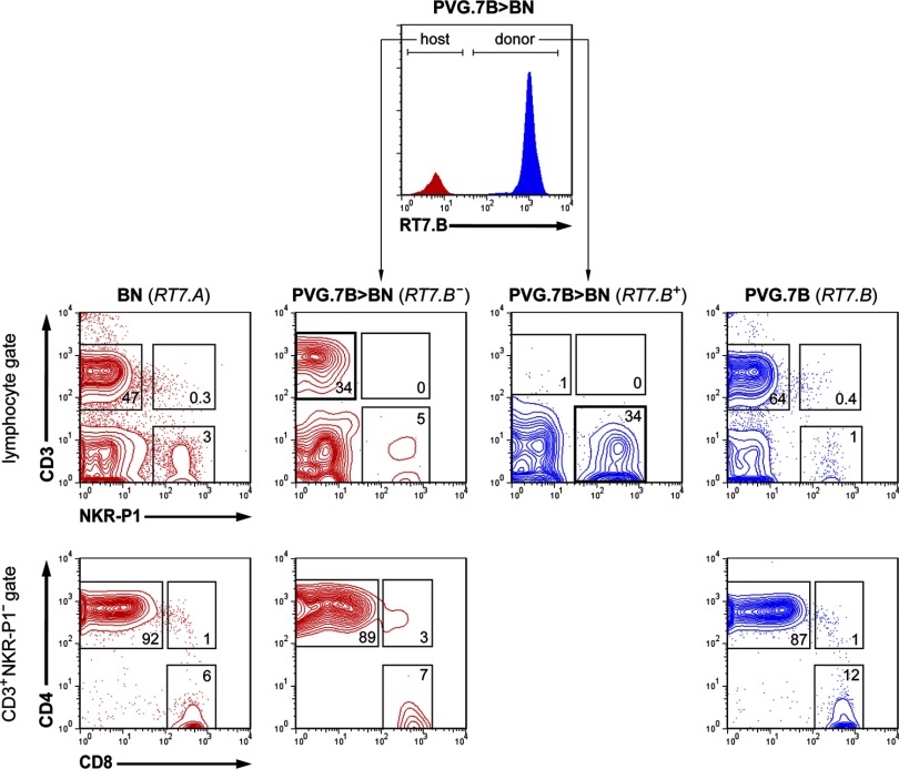
Figure 3. PB composition after BMT.
Representative flow cytometric analyses (five-percentile contour plots) of PB lymphocytes (gated on forward- and side-scatter) from normal BN (far left) and PVG.7B rats (far right) are juxtaposed with the composition of host (RT7.B–) and donor (RT7.B+)-derived cells from BN rats 14 days after allogeneic T cell-depleted BMT and prior to DLI (PVG.7B>BN; cf Fig. 1). T cell (CD3+NKR-P1–), NK cell (CD3–NKR-P1+), and NKT cell (CD3+NKR-P1+) populations (middle row) were gated. Only few T cells derived from PVG.7B donor BM cells were present in the PB of transplanted rats, whereas host T cells and donor NK cells (bold squares) were predominant. T cells were further divided into CD4 (CD4+CD8–), CD8 (CD4–CD8+), as well as “double-positive” (CD4+CD8+) subpopulations (bottom row). Host-derived T cells were mostly CD4+CD8–. Numbers indicate the relative frequencies (% of parent) of the respective gates.

