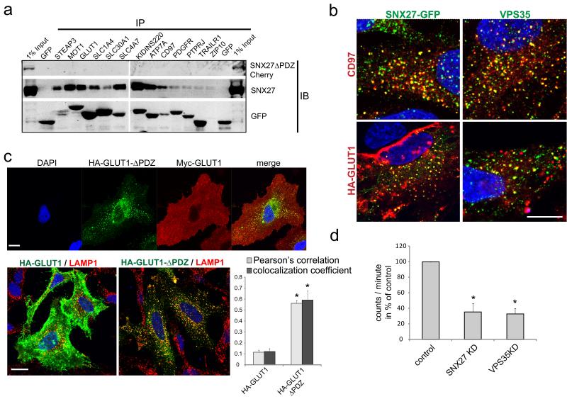
Fig. 3. PDZ interaction motifs at the C-terminus of membrane proteins interact with SNX27 and are required to maintain surface levels.
(a) The cytoplasmic tails of selected proteins from the surface proteome analysis were fused to GFP and analyzed for binding of endogenous SNX27 and mCherry-tagged SNX27ΔPDZ in immunoprecipitation experiments (uncropped blots shown in Fig. S5) (b) Antibody uptake experiments with antibodies against endogenous CD97 and HA-tagged Glut1. After 1h incubation at 37°, internalized Glut1 and CD97co-localized extensively with endogenous VPS35 or GFP-SNX27 on vesicular structures. (c) Upper panel: Immunofluorescent staining of HA-tagged Glut1 full length and Myc-tagged Glut1 with a truncated PDZ binding motif (Glut1-ΔPDZ), lower panel: Colocalization analysis of HA-Glut1 and HA-Glut1ΔPDZ with the lysosomal marker LAMP1. The graph represents the mean of three independent experiments with 10 images each. (n=3, * indicates p<0.05, unpaired t-test; error bars = s.d., individual data points shown in statistics source data, scale bar = 10μm) (d) Uptake assay with radioactive glucose in SNX27 and VPS35 depleted cells. The graph represents the mean of four independent experiments done in quadruplicates. (n=4,* indicates p<0.05, unpaired t-test; error bars = s.e.m., individual data points shown in statistics source data)

