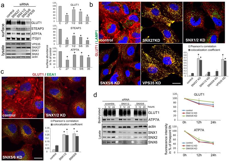
Fig. 4. Suppression of retromer SNX-BAR proteins does not phenocopy SNX27 and VPS35 loss of function but appears to cause trafficking delays.
(a) HeLa cells were transfected with the indicated siRNAs and surface abundance of Glut1, ATP7A and Steap3 was determined by quantitative western blotting (graph represents the mean of 3 (ATP7A), 6 (GLUT1) and 5 (STEAP3) independent experiments, * indicates p<0.05, unpaired t-test; error bars = s.e.m., individual data points shown in statistics source data, uncropped blots shown in Fig. S5) (b) Immunofluorescent colocalization analysis of endogenous Glut1 and LAMP1 in SNX27-retromer and SNX-BAR depleted cells. The graph represents the mean of 30 images acquired in three independent experiments (n=3, * indicates p<0.05, unpaired t-test; error bars = s.d., Scale bar = 10μm, individual data points shown in statistics source data) (c) Colocalization analysis of endogenous Glut1 and the early endosome marker EEA1 in SNX-BAR depleted cells. The graph represents the mean of three independent experiments with 10 images each. (n=3, * indicates p<0.05, unpaired t-test; error bars = s.d., Scale bar = 10μm, individual data points shown in statistics source data) (d) Mean of four degradation assays of Glut1 and ATP7A in SNX-BAR depleted cells (for assay details see Fig. 2D or methods, n=4, * indicates p<0.05, unpaired t-test; error bars = s.e.m., individual data points shown in statistics source data, uncropped blots shown in Fig. S5)

