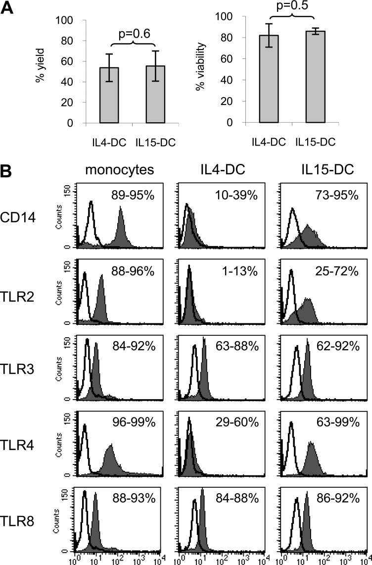
Figure 1. TLRs are differentially expressed by IL4-DCs and IL15-DCs.
Freshly thawed monocytes were labeled immediately for flow cytometric analysis or cultured with GM-CSF + IL-4 or GM-CSF + IL-15 for 3 days to generate IL4-DCs and IL15-DCs, respectively. (A) DCs were harvested, and the percent yield and viability were calculated by trypan blue exclusion. Data represent the mean ± sd of four independent experiments. (B) Cell populations were labeled with indicated anti-human mAb (filled histograms) or appropriate Ig isotype controls (open histogram overlays) and analyzed by multiparameter flow cytometry. For detection of TLR3 and TLR8, cells were fixed and permeabilized prior to labeling with anti-human mAb (filled histograms) or appropriate Ig isotype controls (open histogram overlays) and analyzed by multiparameter flow cytometry. Numbers indicate the range of percent-positive cells. Histograms from one of four independent experiments are shown.

