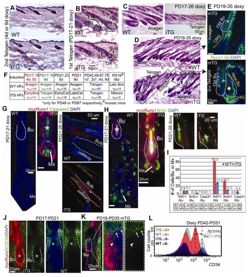
Figure 3. Runx1 expression is sufficient for catagen implementation in bulb cells but requires additional anagen inductive signals to promote stem cell proliferation.

(A-D) Hematoxylin and Eosin (H&E) stained skin sections from doxy-induced mice at stages indicated. WT were littermate controls. Note normal telogen morphology in Runx1iTG and WT in (A). Note Runx1iTG HFs with smaller bulb and lack of hair shaft indicative of earlier anagen, with a DP unenclosed by the epithelial cells in (B) followed by return to telogen with a single hair shaft (*) in (C). (D) HFs in the 16-d induced mosaic Runx1mTG skin showed variable hair cycle stages (dotted rectangles). Corresponding stages are shown in (E), with Runx1 Ab staining (green). Note endogenous Runx1 expression pattern in anagen HF (top) and transgenic pattern (with a positive Bu and interfollicular epidermis) in late catagen/telogen HF (bottom). Note the presence of two hair shafts (*) indicating this HF had been through at least a portion of the 1st adult anagen. White asterisk marks the old shaft (club hair) in the old bulge and yellow asterisk marks the new shaft. (F) Summary of short-term doxy induction and results. (G, H) Staining for markers indicated and quantified in (I) reveal increased BrdU incorporation (2-hr pulse) in Runx1iTG bulge (Bu) and massive apoptosis in the matrix (Mx) and bulb. (J, K) Skin sections from Runx1iTG and Runx1mTG mice show down-regulation of CD34 by immunostaining. Note mosaic expression of mycRunx1 in bulge (Bu) in (K), correlating with lack of CD34 expression. (L) FACS histogram shows overlay of 4 populations isolated from Runx1iTG and WT littermate mouse skin when bulge SCs were at catagen/telogen. Note several fold decrease in CD34 level in CD34+/α6+ Runx1iTG bulge cells compared to WT bulge. See also Fig. S3 and S4.
