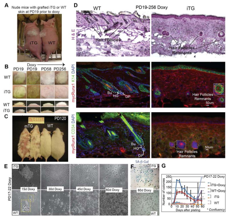
Figure 4. Long-term Runx1 induction in bulge SCs confer a short-lived progenitor cell behavior both in vivo and in vitro.

(A) Grafted newborn Runx1iTG skin onto nude mice, to bypass lethality of Runx1 induction, prior to doxy feeding at the equivalent PD19. (B) Grafted areas on nude mice at times post-induction as indicated. Note permanent hair loss in Runx1iTG skin. (C) Runx1mTG mice induced with doxy for >3 months show permanent hair loss in a patchy manner, as expected from mosaic expression of Runx1. (D) Skin sections from (B) sectioned and stained as indicated show lack of HFs in Runx1iTG skin. Circles show rare remnants of HF cells, which are stained positive for mycRunx1 and K14, but are negative for the SC marker CD34. MycRunx1 staining shows higher dermal background in Runx1iTG than WT, possibly due to increased cellularity in the former. (E-G) Keratinocytes isolated from PD17-PD22 doxy treated mice were plated on irradiated mouse embryonic fibroblast in low Ca2+ E medium. Colonies were counted every week; representative phase contrast images are shown in (E), and the overall counts are shown in (G). See also S6A-B. Note diminished colony size and flat cell morphology indicative of senescence at 60 days in Runx1iTG cells. (F) Endogenous SA-β-Gal staining indicates senescence of Runx1iTG cells by 60 days. See also Fig. S6C.
