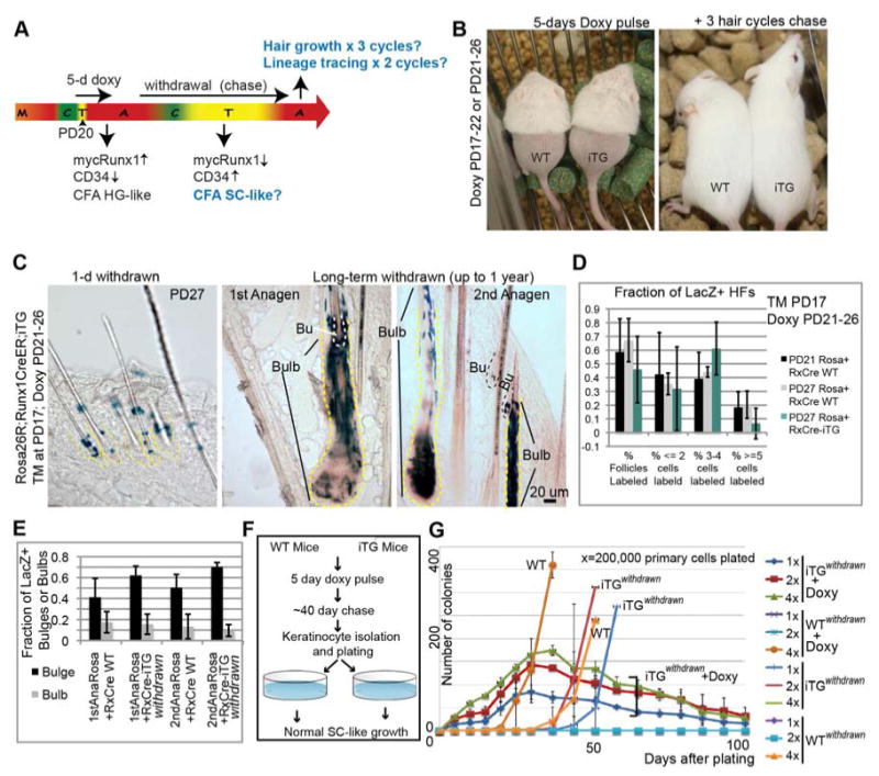
Figure 6. Bulge stem cell fate transition induced by Runx1 is functionally reversible.

(A) Scheme of doxy induction and withdrawal to assess reversibility of Runx1-induced effects on bulge-cell function. (B) Mice on doxy food (left) were gently removed hair on their lower back to monitor hair growth without causing activation of growth by injury. They regrew hair after doxy withdrawal (right) for 3 consecutive cycles (compare with Fig. 4B). (C) Skin sections from Runx1-CreER;Rosa26R;Runx1iTG mice injected with TM at PD17, doxy treated from PD21-PD26, and sacrificed at PD27 (left panel) show localization of LacZ+ cells to the bulge. Middle and right panels show LacZ+ cells in the differentiated HF compartment (bulb) and in the SC compartment (Bu) at the 2 subsequent anagen stages after TM injection. (D, E) LacZ+ HF counts of images collected from all stages in (C), show similar patterns of LacZ contribution to HF regeneration in iTG and WT skin. See also Fig. S7. (F) Scheme of doxy treatment and withdrawal for colony formation assays (CFA) to examine the growth potential (self-renewal ability) of cells that transiently experienced mycRunx1 expression. (G) CFA of cells from (F) show recovery of SC-like behavior in cells from mice in which doxy was withdrawn for several weeks after the initial 5-days of doxy treatment (note iTGwithdrawn vs. WT plots). Keratinocytes from these mice, cultured with doxy upon plating, displayed once again the behavior described previously for the HG (rapid proliferation followed by death; see plots marked iTGwithdrawn +doxy). See also Fig. S7.
