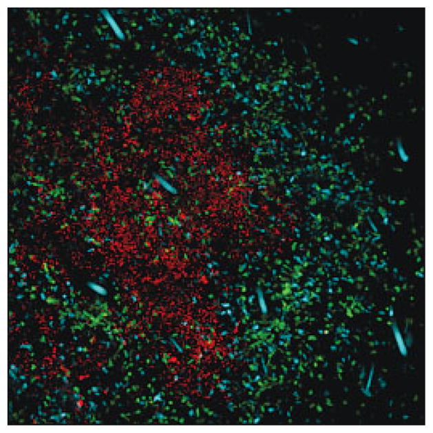
Figure 1.
Foxp3+ Treg accumulate at sites of Leishmania infection. TCR−/− mice were injected with CD4+CD25− T cells from cyan fluorescent protein-expressing transgenic mice and CD4+Foxp3+ Treg from Foxp3-GFP-knock-in mice. Leishmania expressing red fluorescent protein were injected intra-dermally into the ear. This image was taken at 1 month post-infection (red: Leishmania; cyan: CD4+CD25− T cells; green: CD4+Foxp3+ Treg). dysregulation of Treg function requires further analysis.

