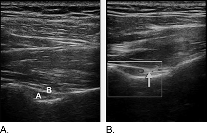
Figure 3.
A: The letter A indicates the area for the suprascapular vessels; the letter B indicates the transverse scapular ligament. B: With application of color Doppler. The suprascapular nerve (indicated by the arrow) can be visualized medial to the pulsation of the suprascapular artery as an oval or round slightly hyperechoic structure.

