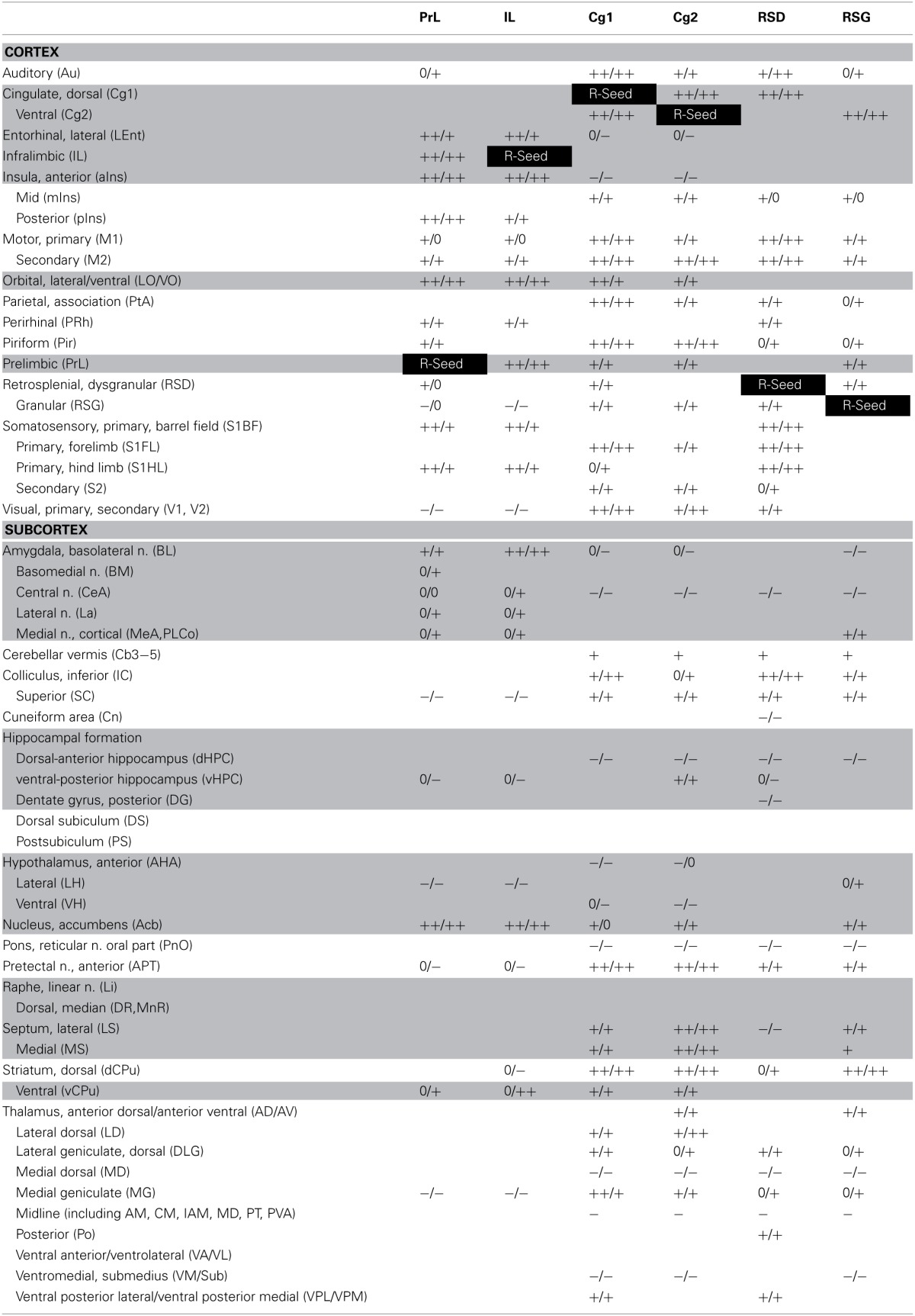
Table 2.
Summary of seed correlation analysis results in the fear-conditioned mice.

Functional connectivity of the cortical midline structures was analyzed using seed correlation for the right prelimbic (PrL), infralimbic (IL), cingulate area 1 (Cg1), cingulate area 2 (Cg2), retrosplenial dysgranular (RSD) and retrosplenial granular (RSG) cortices. Shown are significant left and right (L/R) positive (+) and negative (−) correlations with the seed (P < 0.05, clusters ≥ 100 voxels), with double signs denoting broadly represented correlations. “0” and blank cells denote the absence of significant correlations. Gray shaded cells highlight limbic/paralimbic areas. White text on a black background denotes the seed region.
