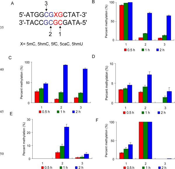
Fig. 2.
Levels of cytosine methylation in different substrates methylated by Dnmt1 at three different CpG sites. (A) The sequence of the duplex DNA employed for the in vitro methylation assay. 1, 2 and 3 indicate the potential methylation sites. The substrates are duplex ODNs containing a 5mC (B), 5hmC (C), 5fC (D), 5caC (E) or 5hmU (F). The data represent the

