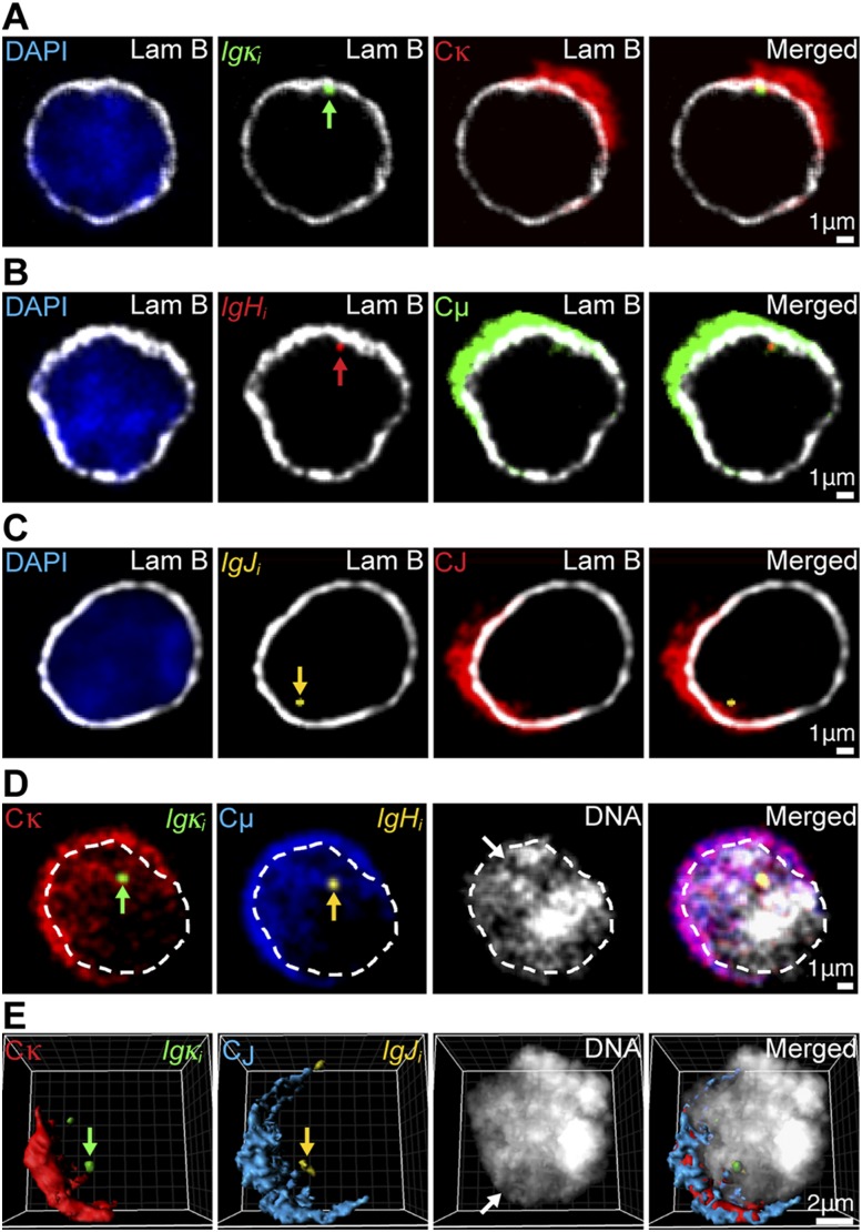
Figure 4.
Transcripts originating from different Ig genes exhibit features of nuclear organization favorable for export to the cytoplasm. (A–C) Representative four-color 3D RNA immuno-FISH images of the same 0.3-μm optical sections of individual plasma cells with peripherally localized transcribing Ig genes. Primary transcripts are indicted by green, red, or yellow arrows. (A) (Blue) DAPI; (white) α-Lam B; (green) Igκi; (red) Cκ. (B) (Blue) DAPI; (white) α-Lam B; (red) IgHi; (green) Cμ. (C) (Blue) DAPI; (white) α-Lam B; (yellow) IgJi; (red) CJ. (D,E) (Red) Cκ; (green) Igκi; (blue) Cμ or CJ; (yellow) IgHi or IgJi; (white) DAPI. White arrows depict interchromatin channels occupied by processed Ig gene transcripts. Primary transcripts of colocalized alleles are indicated by green or yellow arrows. (D) Representative five-color 3D RNA FISH images of the same 0.3-μm optical section of a plasma cell, with interiorly colocalized Ig genes and the nucleus outlined by white dashed lines. (E) Representative reconstructed five-color 3D RNA FISH image of a plasma cell with interiorly colocalized Ig genes.

