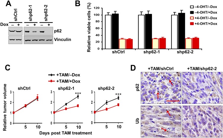
Figure 2.
p62 Knockdown decreases the growth of autophagy-deficient tumors. (A) Various transformed MEFs were treated with 250 nM 4-OHT to delete FIP200 and then cultured for 2 d with or without 1 μg/mL Dox, as indicated. Cell lysates were analyzed by Western blotting for the indicated proteins. (B) Various transformed MEFs (5 × 10⁴) were seeded into six-well plates, and the percentage (normalized to cells under −4-OHT/−Dox conditions) of the number of cells was determined after 5 d in DMEM + 10% FBS culture medium under various conditions, as indicated. (C) Various transformed MEFs were injected subcutaneously into athymic nude mice, all animals (n = 4 each) were treated with TAM by i.p. injection for FIP200 deletion and fed without (black line) or with (red line) Dox-containing food, and single tumor growth was measured at the indicated time points. Data points represent means of fold change and SD in tumor volume relative to 1 d after the last TAM injection. (***) P < 0.001. (D) Representative images of tumor sections harvested from the recipient mice at the final time point and analyzed by immunohistochemistry with anti-p62 and anti-ubiquitin (anti-Ub) antibodies. Arrows mark p62-positive and ubiquitin-positive aggregates. Bar, 100 μm.

