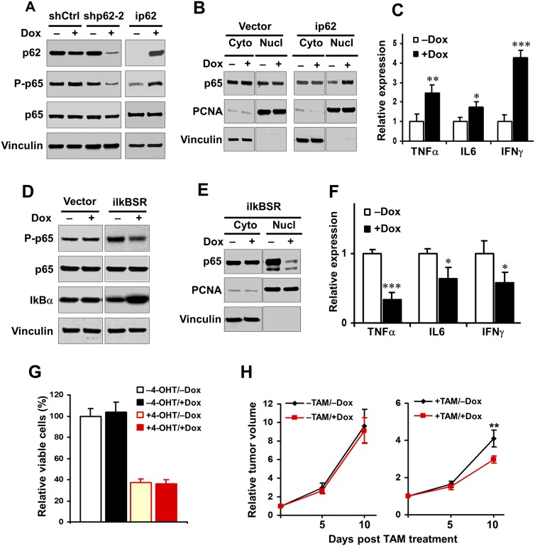
Figure 4.
p62 regulates NF-kB activation, and inhibition of NF-kB impairs the tumor growth under autophagy-deficient condition. (A) Various transformed MEFs were treated with 250 nM 4-OHT to delete FIP200 and then cultured for 2 d with or without 1 μg/mL Dox to induce p62 knockdown (left four lanes) or ectopic p62 expression (right two lanes), as indicated. Cell lysates were analyzed by Western blotting for the indicated proteins. (B) Various transformed MEFs were treated with 250 nM 4-OHT to delete FIP200 and then cultured for 2 d with or without 1 μg/mL Dox, as indicated. Cytoplasmic (Cyto) and nuclear (Nucl) fractions were then prepared and analyzed by Western blotting for the indicated proteins. (C) ip62 cells were treated with 250 nM 4-OHT for deletion of FIP200 and then cultured for 2 d with or without 1 μg/mL Dox. RNA was isolated from cells and subjected to the analysis by quantitative RT–PCR (qRT–PCR) to detect the expression of NF-kB target genes, as indicated. The mean ± SD of relative levels (normalized to no Dox treatment) is shown. (D) Various transformed MEFs were treated with 4-OHT to delete FIP200 and then cultured overnight with or without 1 μg/mL Dox, as indicated. Cell lysates were analyzed by Western blotting for the indicated proteins. (E) iIkBSR cells were treated with 250 nM 4-OHT to delete FIP200 and then cultured for 2 d with or without 1 μg/mL Dox, as indicated. Cytoplasmic (Cyto) and nuclear (Nucl) fractions were prepared and analyzed by Western blotting for the indicated proteins. (F) iIKBSR cells were treated with 250 nM 4-OHT for deletion of FIP200 and then cultured for 2 d with or without 1 μg/mL Dox. RNA was isolated from cells and subjected to the analysis by qRT–PCR to detect the expression of NF-kB target genes, as indicated. The mean ± SD of relative levels (normalized to no Dox treatment) is shown. (G) iIkBSR cells (5 × 10⁴) were seeded into six-well plates, and the percentage (normalized to cells under −4-OHT/−Dox conditions) of the number of cells was determined after 5 d in DMEM + 10% FBS culture medium under various conditions, as indicated. (H) iIkBSR cells were injected subcutaneously into athymic nude mice, animals (n = 4 each) were treated with or without TAM by i.p. injection for FIP200 deletion and fed without (black line) or with (red line) Dox-containing food, and single tumor growth was measured at the indicated time points. Data points represent means of fold change and SD in tumor volume relative to 1 d after the last TAM injection. (*) P < 0.05; (**) P < 0.01; (***) P < 0.001.

