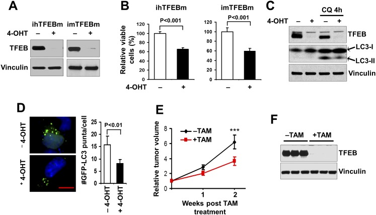
Figure 5.
Enhanced autophagy by ectopic expression of the TFEBS142A mutant correlates with increased tumor growth. (A) ihTFEBm (left) or imTFEBm (right) cells were treated with 4-OHT for 2 d to delete the ectopic floxed TFEBS142A mutant TFEB and then cultured overnight in growth medium, as indicated. Cell lysates were analyzed by Western blotting for the indicated proteins. (B) ihTFEBm (left) or imTFEBm (right) cells (5 × 10⁴) were seeded into six-well plates, and the percentage (normalized to cells under −4-OHT conditions) of the number of cells was determined after 6 d in culture medium under various conditions, as indicated. (C) imTFEBm cells were treated with 4-OHT as described in A and then replated in growth medium with (right two lanes) or without (left two lanes) 50 μM chloroquine (CQ) for 4 h. Cell lysates were analyzed by Western blotting for the indicated proteins. (D) ihTFEBm cells were infected by recombinant retroviruses encoding GFP-LC3 and then treated with 250 nM 4-OHT to delete floxed TFEBS142A. The cells were then replated and cultured in serum-starved medium overnight. Representative images (left panels) and the mean ± SD (right graph) of GFP-LC3 puncta (green) are shown, respectively. Slides were counterstained with DAPI (blue). Bar, 5 μm. (E) imTFEBm tumor cells were injected into the mammary fat pads of nude mice. Animals (n = 5 each) were treated with vehicle control (black line) or TAM (red line) by i.p. injection, and single tumor growth was measured at the indicated time points. Data points represent means of fold change and SD in tumor volume relative to 1 d after the last TAM injection. (***) P < 0.001. (F) Lysates were prepared from mammary tumors of recipient mice and then analyzed by Western blotting for the indicated proteins.

