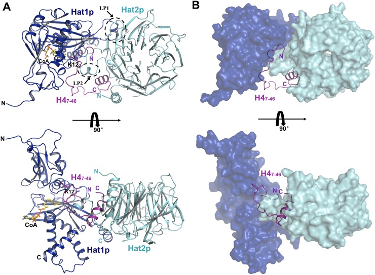
Figure 1.
Crystal structure of yeast Hat1p/Hat2p/H4 peptide/CoA. (A) Overall structure of the Hat1p/Hat2p/H4 peptide/CoA complex. The protein structure is shown in the cartoon, and CoA is shown in stick representation, with Hat1p, Hat2p, and H4 peptide colored in blue, cyan, and purple, respectively. (B) H4 peptide in the structure of the Hat1p/Hat2p/H4 peptide/CoA complex. Hat1p and Hat2p are shown in surface mode, with Hat1p colored blue, and Hat2p colored cyan.

