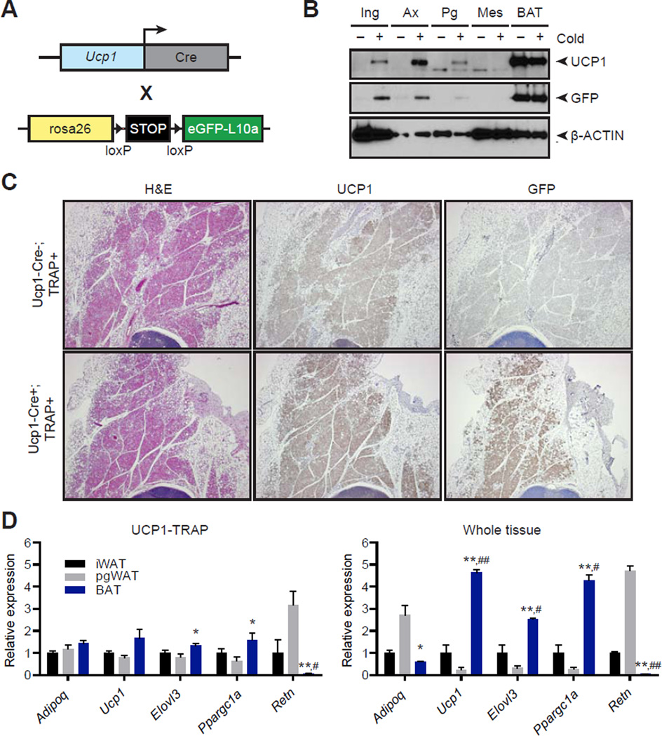
Fig. 1. Characterization of the UCP1-TRAP mouse.
(A) Schematic of the cross to generate UCP1-TRAP mice. A BAC transgenic expressing Ucp1-Cre was crossed with a TRAP mouse expressing a fusion protein of eGFP and the ribosomal subunit L10a (eGFP-L10a, referred to as the TRAP allele) under the control of a lox-stop-lox cassette in the rosa26 locus. (B) Western blot analysis of UCP1, GFP, and β-actin loading control from various fat depots in 6-week old UCP1-TRAP mice at room temperature (−) or after two weeks cold exposure (+) at 4°C. Ing, inguinal WAT; Ax, axillary WAT; Pg, perigonadal WAT; Mes, mesenteric WAT. (C) Representative H&E, UCP1, and GFP immunohistochemistry from 6-week old male UCP1-TRAP (Cre-positive, TRAP-positive) or control mice (Cre-negative, TRAP-positive) after two weeks cold exposure at 4°C. (D) Relative mRNA expression of the indicated genes from various fat depots in UCP1-TRAP samples (left) or whole tissues (right). Tissues were harvested from 6-week old female mice after two weeks cold exposure at 4°C. Data are presented as means ± standard error; n = 3–5/group. *, ** p < 0.05 or < 0.01, respectively, in BAT versus pgWAT samples; #, ## p < 0.05 or < 0.01, respectively, in BAT versus iWAT samples.

