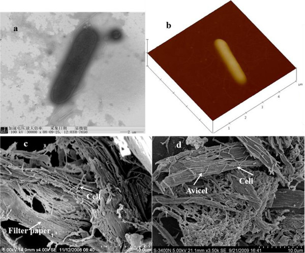
Figure 3.

Micrographs of T. thermosaccharolyticum M18. (a) Transmission electron microscopy (TEM) micrograph, (b) atomic force microscopy (AFM) micrograph, and scanning electron microscopy (SEM) images of strain M18 cells associated with the cellulose fibers supplied in the filter paper culture (c) and Avicel culture (d).
