Figure 3.

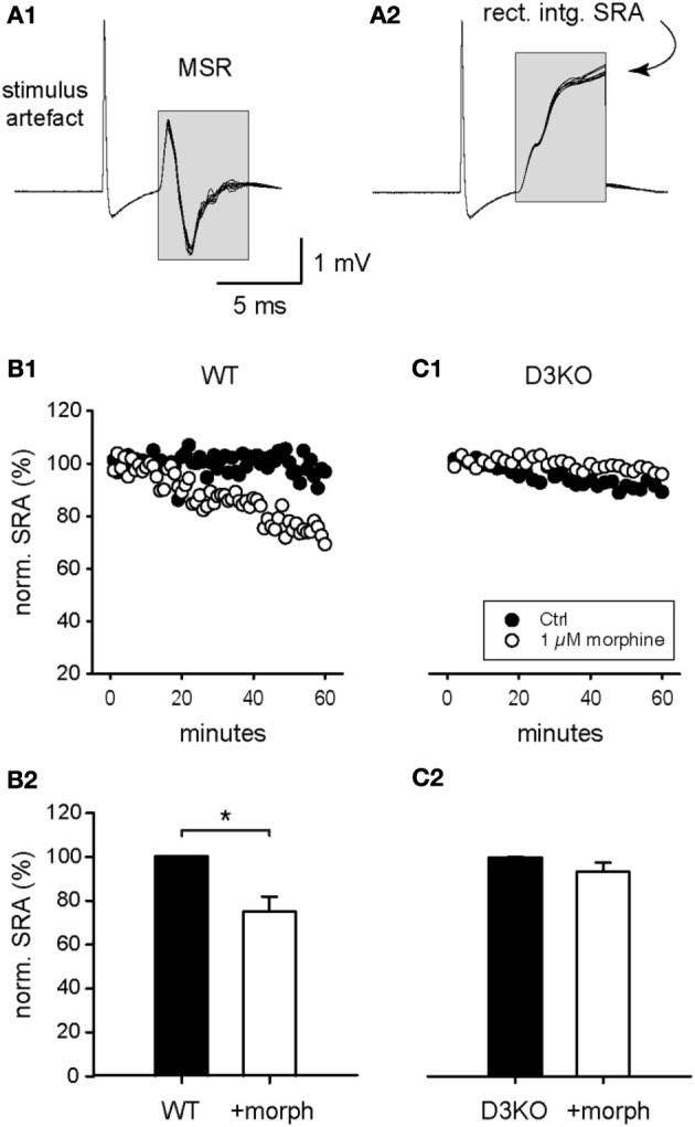
Modulation of spinal reflex amplitudes (SRAs) by morphine. (A1) Example of a reflex response recorded from ventral roots (10 consecutive sweeps superimposed). Stimulation of a dorsal root (at stimulus artifact) induced a monosynaptic reflex response (MSR) in the corresponding ventral root. The boxed region identifies the epochs chosen to calculate the amplitude of the spinal reflex amplitude (SRA) represented in (A2). (A2) The rectification and subsequent integration of the epochs identified in (A1) led to the signal values highlighted in the boxed region. The final maximal values of each epoch were used as a measurement of the SRA for each sweep, and used in the subsequent analysis steps represented in panels (B,C), and Figures 4, 5. (B1) Typical time course of control SRAs in WT before and during bath-application of 1 μM morphine (control: black symbols; morphine: white symbols). During morphine, reflex amplitudes gradually decreased in amplitude. (B2) Normalized SRAs in WT (n = 6). Morphine led to a significant reduction in SRA. *p = 0.004. (C1) Typical time course of control SRAs in D3KO before and during bath-application of 1 μM morphine (control: black symbols; morphine: white symbols). (C2) Normalized SRAs in D3KO (n = 12). In these animals, morphine had no significant effect on SRA.
