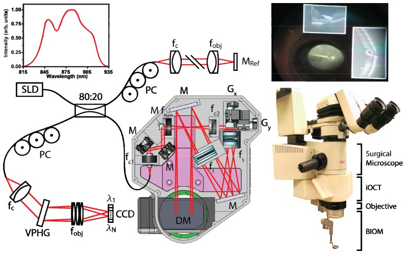
Fig. 1.
Optical schematic and mechanical design of the microscope-integrated iOCT system (Media 1 (3.3MB, AVI) ). The system includes an electrically tunable lens to provide optimal focus for both anterior and posterior segment imaging while maintaining parfocality with the microscope oculars. OCT images were acquired with a modified commercial 36 kHz SDOCT engine. Inset photo shows surgical microscope ocular view with HUD overlaid live OCT cross-sections adjacent to conventional microscope view of retina. CCD, line-scan camera; DM, dichroic mirror; f, collimating, objective, scan, tube, and electrically tunable lenses; G, galvanometer scanners; M, reference and fold mirrors; PC, polarization controller; VPHG, grating.

