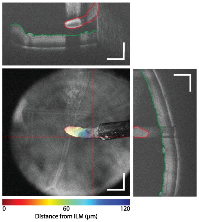
Fig. 4.

Surgical guidance using iOCT feedback. The surface of the membrane scraper (red) and ILM (green) were segmented on individual cross-sectional images (dotted line). An axial position difference is overlaid as a colormap on the en face view to provide integrative visualization of three-dimensional data of the membrane scraper position relative to the surface of the retina. Scale bar: 500 µm.
