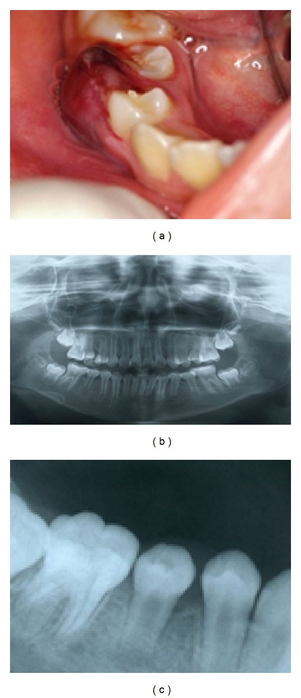
Figure 1.

(a) Preoperative intraoral photograph showing a gingival abscess in the mandibular right second premolar. (b) Panoramic X-ray showing extensive radiolucency in the periradicular region in the mandibular right second premolar compared with the mandibular left second premolar. (c) X-ray showing an immature open apex and enlargement of the periodontal ligament space and extensive radiolucency in the periradicular region in the mandibular right second premolar.
