Figure 4.

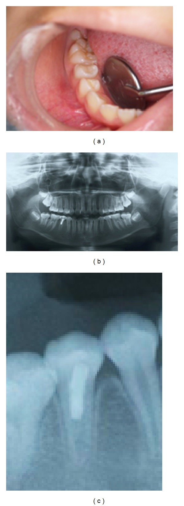
(a) Intraoral photograph showing no abnormalities of gingiva. (b) Panoramic X-ray showing the formation of a dentin bridge and thickening of the canal walls in the mandibular right second premolar. (c) Panoramic X-ray showing the formation of a dentin bridge and thickening of the canal walls and establishment of the periodontal ligament space and lamina dura in the mandibular right second premolar.
