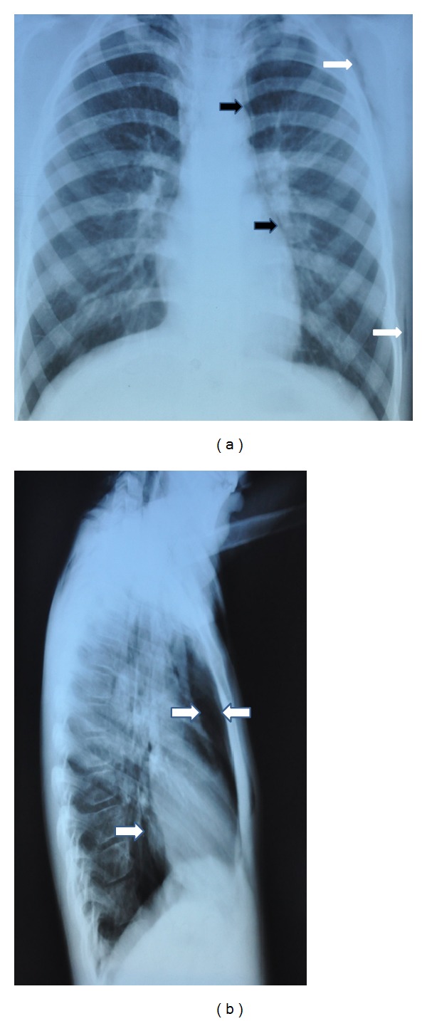
Figure 1.

(a) Anteroposterior chest radiograph showing thin radiolucent line outlining aortic root and left heart border (black arrows) and subcutaneous emphysema (white arrows). (b) Lateral chest radiograph showing retrosternal emphysema (between arrows) and radiolucent line outlining posterior border of the heart.
