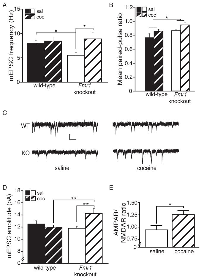
Fig. 5. FMRP opposes cocaine-induced changes in functional synapse number and synaptic strength.
(A) Similar to observations for overall spine density, the frequency of mEPSC events was observed to be uniquely susceptible to COC treatment (5 d, 15 mg/kg + 24 hrs) in Fmr1KO mice (n, cells=11 KO SAL, 16 KO COC, 13 WT SAL, 15 WT COC). Additionally, a significant basal decrease in mEPSC frequency was present in Fmr1 KO compared to WT mice (planned comparison). (B) Increased paired-pulse ratio (PPR) (i.e., decreased presynaptic release) was seen in Fmr1 KO compared to WT mice (inter-stimulus interval=100 msec; n, cells=9 KO SAL, 14 KO COC, 7 WT SAL, 11 WT COC). A trend was also present for COC to decrease presynaptic release. (C) Representative traces of mEPSCs from each group. (D) An overall significant interaction was observed for mEPSC amplitude, which was uniquely increased by COC treatment in Fmr1 KO mice and, among COC-treated mice only, KO had higher amplitude than WT. (E) COC also increased the AMPAR/NMDAR ratio in Fmr1 KO MSNs (n, cells=7 SAL, 11 COC). Asterisks in A & D indicate significant follow-up Bonferroni post-hoc tests, except planned comparison (Ind. Samples T-test). (Scale bar=10 pA/100 msec; * p<0.05; data shown are mean±S.E.M.; see also Table S1 & Fig. S5.)

