Figure 2.
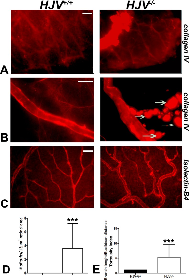
Evidence of neovascularization in Hjv−/− mouse retina. Retinal flat-mounts were stained with collagen IV to label retinal vasculature. (A) Superficial vascular mass was noted at the periphery of the retina in Hjv−/− mice. (B) Blood vessels in Hjv−/− mice showed marked tortuosity with vascular masses (arrows) extending out of the vessel wall compared with normal appearing vasculature in wild-type mouse retina. (C, D) Isolectin-B4–stained retinal flat-mounts were used to count capillary tufts per a given retinal area and to quantify the vessel tortuosity. Scale bar: 20 μm. Data are representative of the experiments performed in three wild-type mice and three Hjv−/− mice.

