Figure 3.
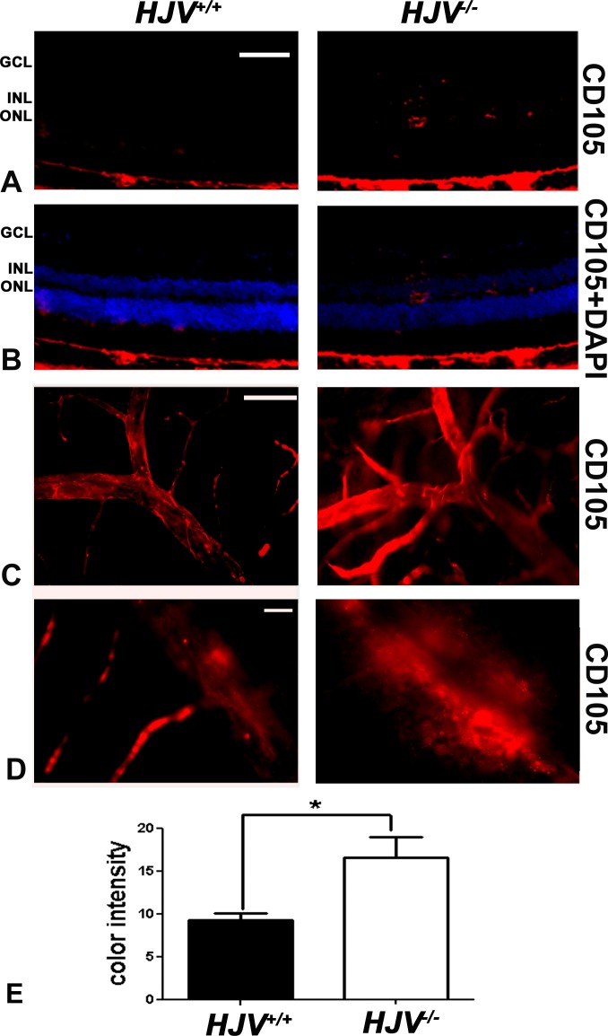
Increased expression of endoglin (CD105) in Hjv−/− mouse retinas. (A, B) Fluorescent immunodetection of endoglin (red), a marker of neovascularization, was performed in retinal cryosections of Hjv+/+ and Hjv−/− mice; DAPI (blue) was used to label nuclei. There was minimal detection of endoglin in Hjv+/+ mouse retinas whereas endoglin levels were markedly increased in Hjv−/− mouse retinas, particularly where the blood vessels are predominant (GCL, inner plexiform layer, and INL). Scale bar: 50 μm. (C, D) Retinal flat-mounts were stained for endoglin and assessed at low magnification (C) and high magnification (D). The retinas from Hjv−/− mice showed markedly branching, tortuous, dilated blood vessels with increased endoglin staining compared with normal appearing and less endoglin-stained retinas from Hjv+/+ mice. Scale bar: 50 μm (C) and 20 μm (D). (E) Quantification of the data obtained from metamorphic analysis of the color intensity of the retinas stained with CD105 in (C) (*P < 0.05, n = 3 mice for each genotype).

