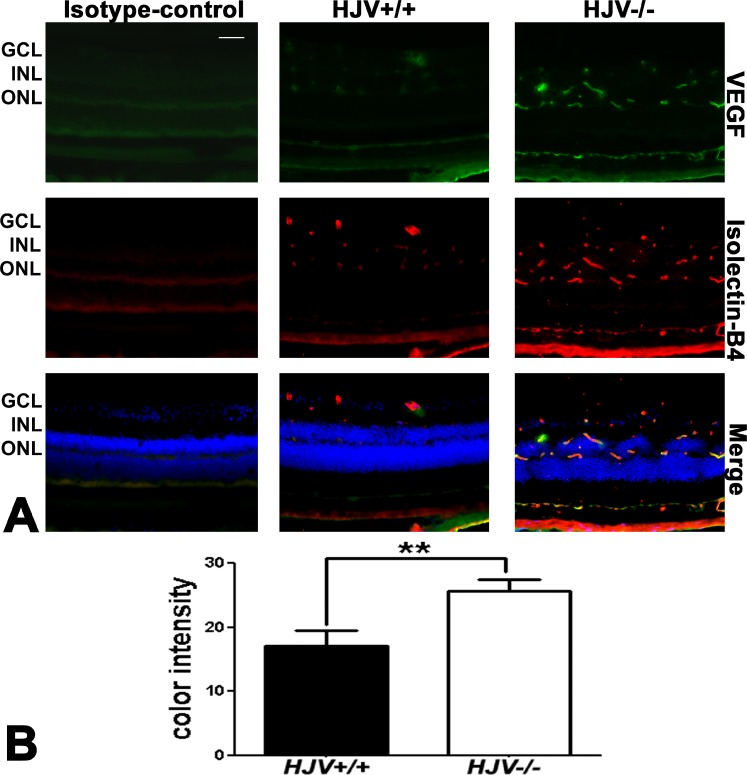
Figure 4.
Increased expression of VEGF in Hjv−/− mouse retinas. (A) Retinal cryosections from Hjv+/+ and Hjv−/− mice were immunostained for VEGF (green), a marker of neovascularization, and isolectin-B4 (red), a marker of blood vessels. The levels of VEGF were markedly increased in the Hjv−/− mouse retinas compared with wild-type (Hjv+/+) mouse retinas. The far left panel is a negative control in which an isotype antibody (IgG1 was used instead of the primary antibody, but with the secondary antibody). Scale bar: 50 μm. (B) Quantification of the intensity levels of VEGF immunofluorescence. **P < 0.01, n = 3 mice for each genotype; Scale bar: 50 μm.

