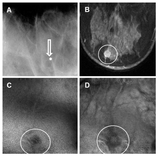
Figure 5.

Images from case 3. (A) Mammogram shows only a marker (arrow) placed on the site of the palpable mass (no mass visible in image). (B) Magnetic resonance image shows the lesion (circle). (C-D) Vibro-acoustography images at a 2.5-cm depth (C) and 3.0-cm depth (D) show the lesion (circles).
