
Figure 3.
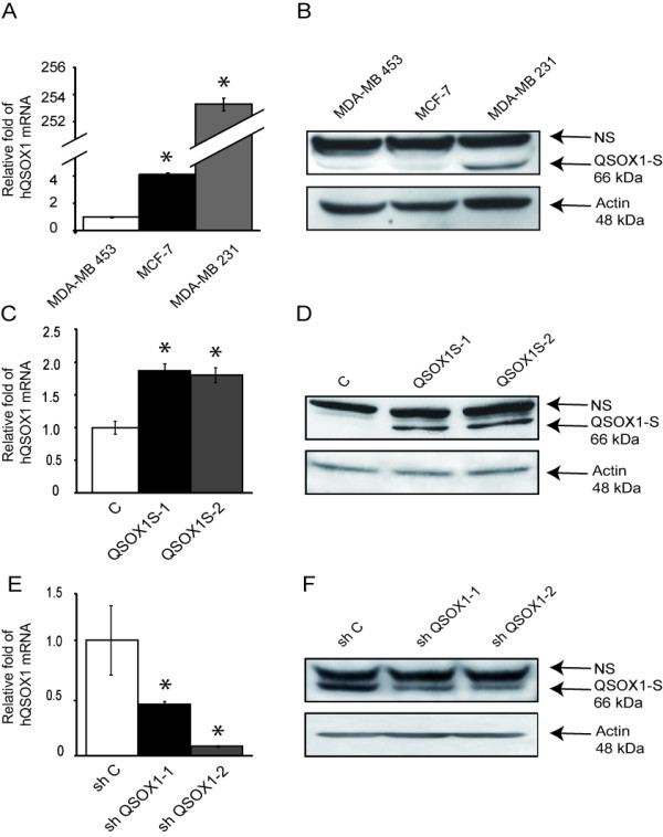
Analysis of QSOX1 expression in breast cancer cell lines. (A, C, E) After reverse transcription, relative QSOX1 mRNA expressions were determined by qPCR (C: MCF-7; E: MDA-MB-231). H3B-2 mRNA was used for normalization. Data are means ± S.D. of three independent experiments. *P <0.05, compared to control (Wilcoxon test). (B, D, F) Western blot analysis of QSOX1 expression. Total proteins (50 μg) were separated on SDS-PAGE and transferred to PVDF membranes (D: MCF-7; F: MDA-MB-231). Proteins were detected using anti-QSOX1 and anti-actin antibodies. NS, Non Specific.