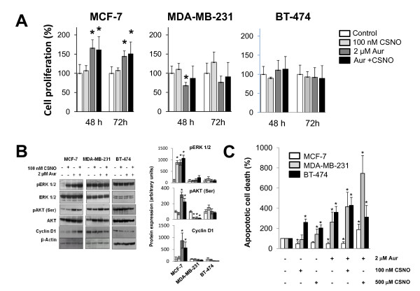
Figure 2.

Mild nitrosative stress increases cell proliferation and survival of MCF-7 cells. (A) Cells were exposed to the indicated treatments and cell proliferation was determined after 48 or 72 hours. Cell proliferation expressed as the percentage of untreated cells. (B) Cells were exposed for 6 hours to the indicated treatments and phosphorylation of Akt and Erk1/2 and cyclin D1 levels was determined by western blot using the corresponding specific antibodies. The corresponding densitometric analyses of the protein bands detected in the immunoblots and normalized to the signal of β-actin are also shown. Data are means ± standard error of the mean of three independent experiments. (C) Cells were exposed to the indicated treatments and apoptotic cell death was determined. The percentage of apoptotic cells in controls was 7.4 ± 1.74, 2.4 ± 0.94 and 2.2 ± 1.04 for MCF-7 cells, MDA-MB-231 cells and BT-474 cells, respectively. Apoptotic cell death expressed as the percentage of untreated cells. *P < 0.05, compared with untreated. #P < 0.05, compared with S-nitrosocysteine (CSNO) treated. Aur, auranofin.
