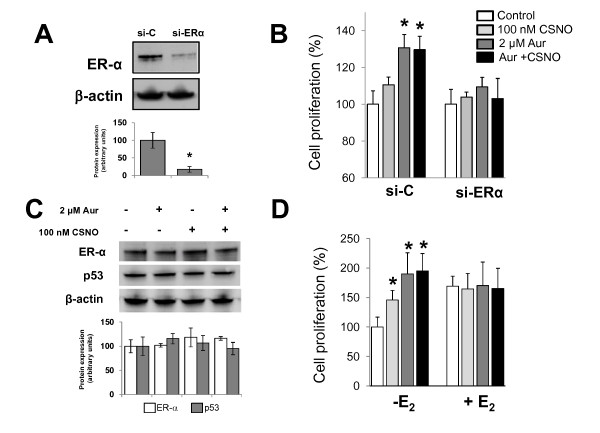
Figure 4.

Proliferative effect of mild nitrosative stress abolished by ERα silencing and increased by estrogen deprivation. (A) Cells were transiently transfected with scrambled siRNA (si-C) or specific si-ERα as described in Materials and methods and ERα expression was analyzed by immunoblotting in whole cell lysates. The corresponding densitometric analysis of the protein band detected and normalized to the signal of β-actin in the immunoblot is also shown. Data are means ± standard error of the mean of three independent experiments. *P < 0.05, compared with cells transfected with si-C. (B) Cells were transiently transfected with si-C or si-ERα, treated as indicated, and cell proliferation was determined after 48 hours of treatment. Cell proliferation expressed as the percentage of untreated cells. *P < 0.05, compared with untreated. (C) Cells were subjected to the indicated treatments and ERα and p53 expression was analyzed by immunoblotting. The corresponding densitometric analysis of the protein band detected and normalized to the signal of β-actin in the immunoblot is also shown. Data are means ± standard error of the mean of three independent experiments. *P < 0.05, compared with untreated cells. (D) MCF-7 cells were maintained in phenol red-free medium with charcoal-stripped serum for 24 hours before exposure to the indicated treatments. Cell proliferation expressed as the percentage of untreated cells. *P < 0.05, compared with untreated. Aur, auranofin; CSNO, S-nitrosocysteine; E2, 17β-estradiol; ER, estrogen receptor.
