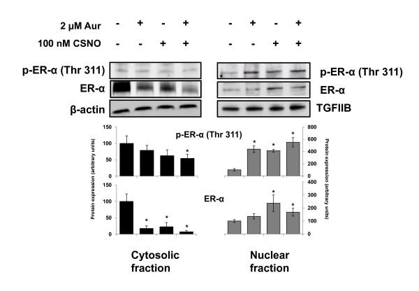
Figure 6.

Alteration of ERα subcellular localization is associated with its phosphorylation at Thr311. Estrogen-deprived MCF-7 cells were exposed to the indicated treatments for 6 hours and, after separation of cytosolic and nuclear fractions, the expression of ERα and p-ERα (Thr311) was analyzed by immunoblotting. Immunodetection of β-actin and TGFIIB were included as loading controls for cytosolic and nuclear fractions, respectively. The corresponding densitometric analyses of the protein bands detected in the immunoblots and normalized to the signal of β-actin or TFIIB are also shown. Data are means ± standard error of the mean of three independent experiments. *P < 0.05, compared with untreated cells. Aur, auranofin; CSNO, S-nitrosocysteine; ER, estrogen receptor; TGF, transforming growth factor.
