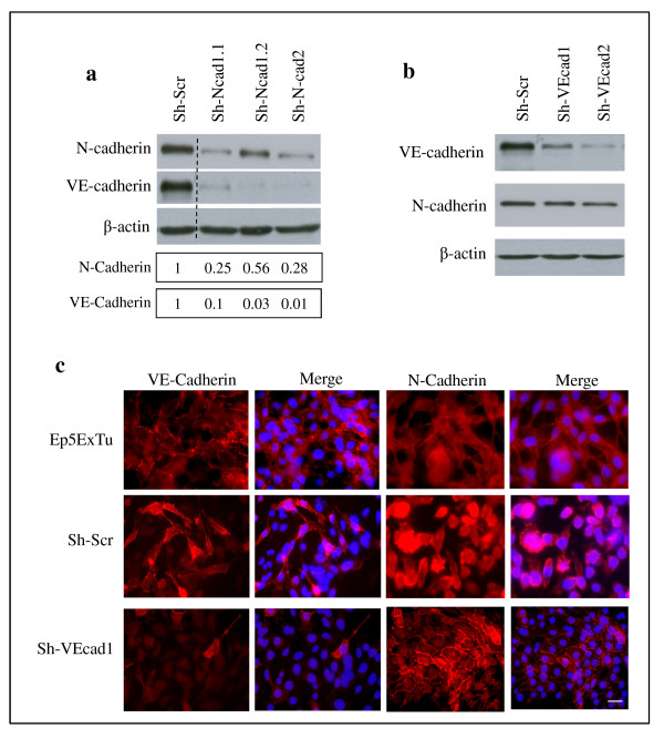
Figure 2.

Neural (N)-cadherin regulates vascular endothelial (VE)-cadherin expression in Ep5ExTu cells. (a) N-cadherin silencing in Ep5ExTu cells transduced either with a scrambled shRNA-containing virus (Sh-Scr) or with Sh-N-cadherin virus (Sh-N-cad1.1, Sh-N-cad1.2 and Sh-N-cad2). The bars represent the relative N-cadherin or VE-cadherin protein levels in Sh-N-cadherin cell lines versus the Sh-Scr cell line, as determined by western blot analysis. (b) VE-cadherin protein expression was examined by western blot in Ep5ExTu cells that were transduced with the VE-cadherin shRNA (Sh-VE-cad-1 and Sh-VE-cad-2) and in the Sh-Scr cell lines. β-actin was detected as loading control. (c) Immunofluorescence staining for VE-cadherin or N-cadherin in Sh-VE-cad-1 and Sh-Scr cells. Ep5ExTu cells were used as a positive control for the localization of VE-cadherin and N-cadherin. Nuclear staining with DAPI is also shown. Bar, 60 μm.
