Figure 3.

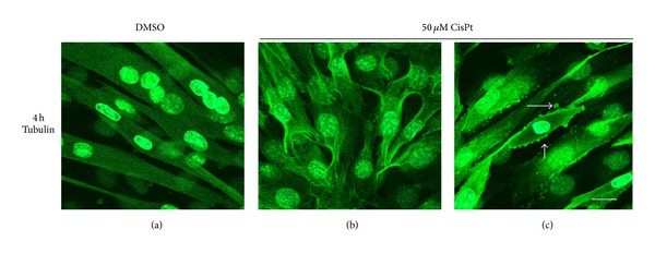
Alpha-tubulin distribution in C2C12 myotubes exposed to CisPt 50 μM for 4 h. Note the anomalous organization of the microtubular network (b)-(c) versus DMSO exposed controls (a). Bar = 10 μm.

Alpha-tubulin distribution in C2C12 myotubes exposed to CisPt 50 μM for 4 h. Note the anomalous organization of the microtubular network (b)-(c) versus DMSO exposed controls (a). Bar = 10 μm.