
Figure 6.
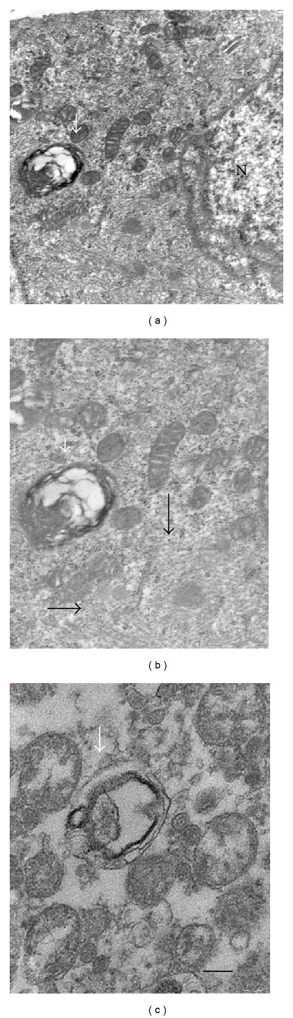
Ultrastructure of C2C12 myotubes challenged with 50 μM CisPt for 24 h in the presence of Tau. (a) Note mitochondria and autophagolysosome around nucleus (N); (b) at higher magnification, autophagosomes, well-organized microtubules (white arrows), and mitochondria with regular cristae (asterisk); (c) destroyed mitochondrion inside an autophagic vacuole (dark arrow) in the culture medium together with vesicles and cellular debris. Bar = 200 nm.