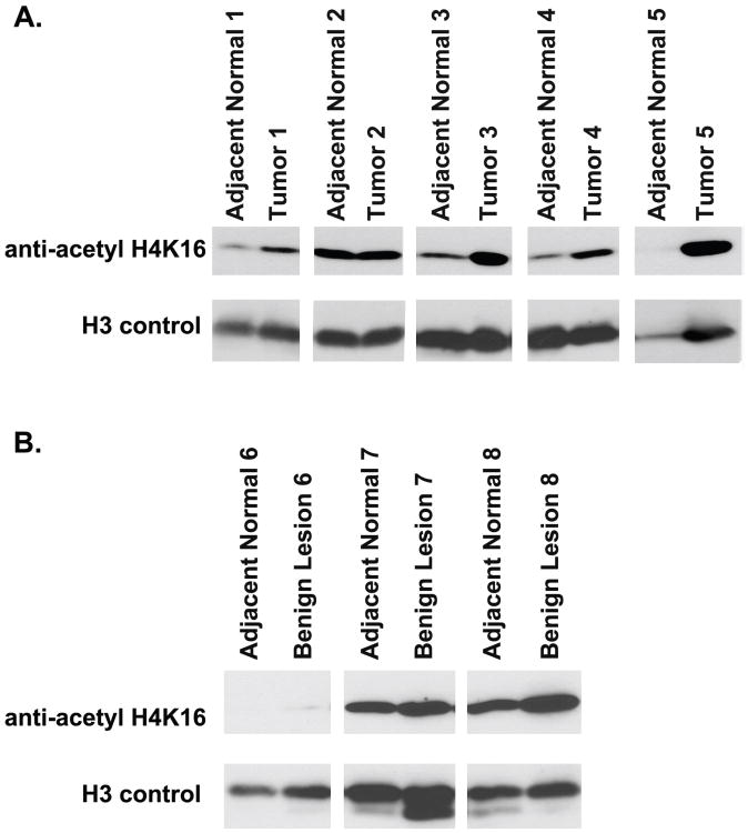
Figure 5.
Western blot analysis of H4K16 acetylation in lung tissue. A. Five paired tumor and adjacent normal lung tissues. Four (1, 3, 4, and 5) samples show increased H4K16 acetylation B. Three paired benign nodules and adjacent normal lung tissue. The paired samples have similar levels of H4K16 acetylation.

