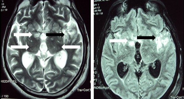
Figure 2.

Fluid attenuated inversion recovery (FLAIR) (Right) and T2-weighted (Left) images of magnetic resonance imaging (MRI) of brain. Bilateral multiple nodular lesions in basal ganglia and thalamus (white arrows). Ill defined area of signal intensity change in the left temporoparietal region (black arrows).
