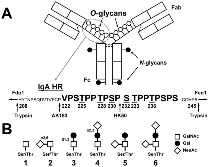
Figure 1. Structure of IgA1 and the hinge-region (HR) amino-acid sequence.
(A) Monomeric IgA1 and its HR with nine possible sites of O-glycan attachment and the Fc portion of heavy chain with two N-glycans. Underlined serine (S) and threonine (T) residues in HR are frequently glycosylated [26], [30], [31]. Arrows show cleavage sites of trypsin and two IgA-specific proteases (from Clostridium ramosum AK183 and Haemophilus influenzae HK50). (B) O-glycan variants of circulatory IgA1∶1, Tn antigen; 2, sialyl-Tn antigen; 3, T antigen; 4, α3-sialyl-T antigen; 5, α6-sialyl-T antigen; 6, disialyl-T antigen. Abbreviations: GalNAc, N-acetylgalactosamine; Gal, galactose; NeuAc, N-acetylneuraminic acid.

