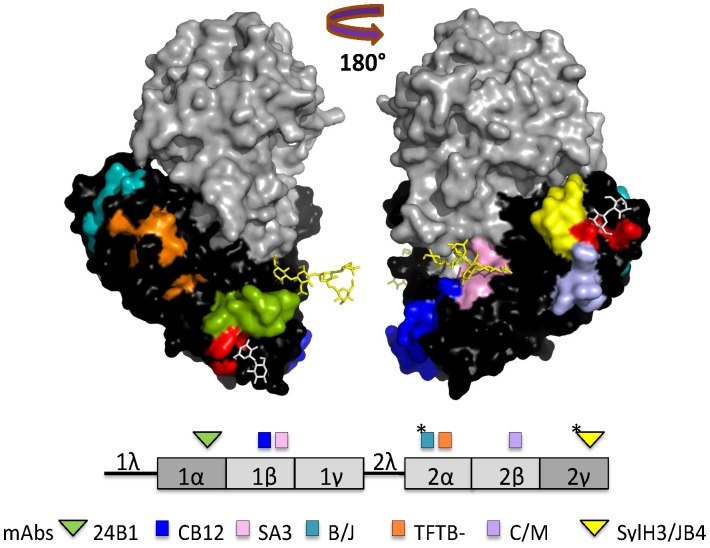
Figure 1. Previously identified and characterized epitopes on RTB recognized by neutralizing and non-neutralizing mAbs.
X-ray crystal structure of ricin holotoxin visualized using PyMOL and based on PDB file 2AAI [55]. RTA (grey), RTB (black), ricin’s N-linked mannose side chains (yellow sticks) and lactose moieties (white sticks) are shown in the upper panel. Confirmed and putative epitopes (*) recognized by neutralizing (triangles) and non-neutralizing (squares) RTB-specific mAbs are color-coded on the holotoxin structure to match RTB’s linear subdomain organization in the lower panel. This figure was modified from an earlier version [26].

