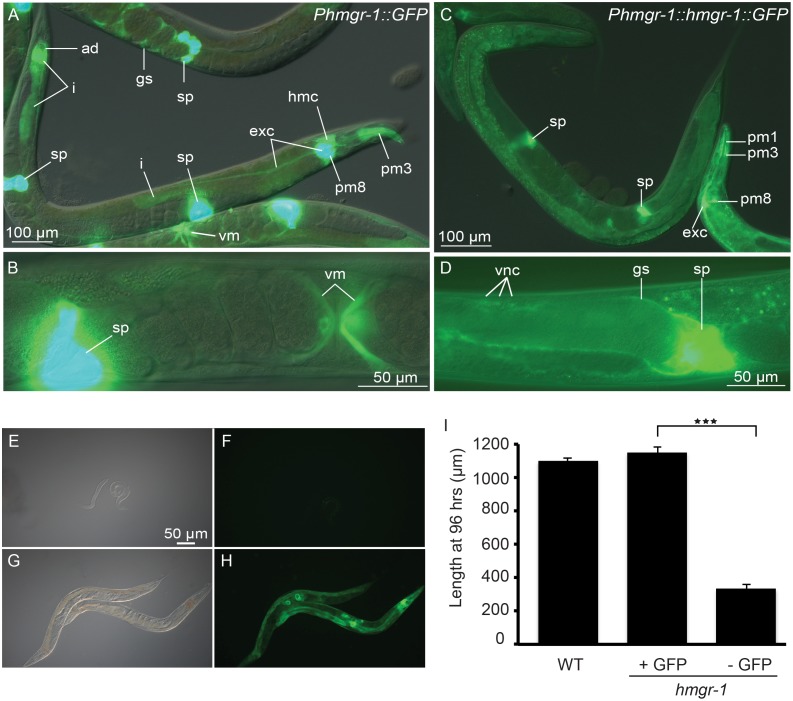
Figure 6. Several tissues express hmgr-1 reporters.
Expression of the Phmgr-1::GFP transcriptional reporter (A–B) and Phmgr-1::HMGR-1::GFP translational reporter (C–D). Structures labeled are as follows: ad (anal depressor), exc (excretory canal), gs (gonad sheath), hmc (head mesodermal cell), i (intestine), pm1, pm3 and pm8 (pharyngeal muscles 1, 3 and 8), sp (spermatheca), vm (vulva muscles), vnc (ventral nerve cord). (E–F) and (G–H) respectively show GFP-negative and GFP-positive progeny from hmgr-1(4368); Ex[Phmgr-1::HMGR-1::GFP rol-6] transgenic animals grown on normal plates, i.e. without exogenous mevalonate; the GFP-positive progeny grow as well as wild-type animals while the GFP-negative progeny do not grow (I). ***p<0.001 using Student’s t-test.

