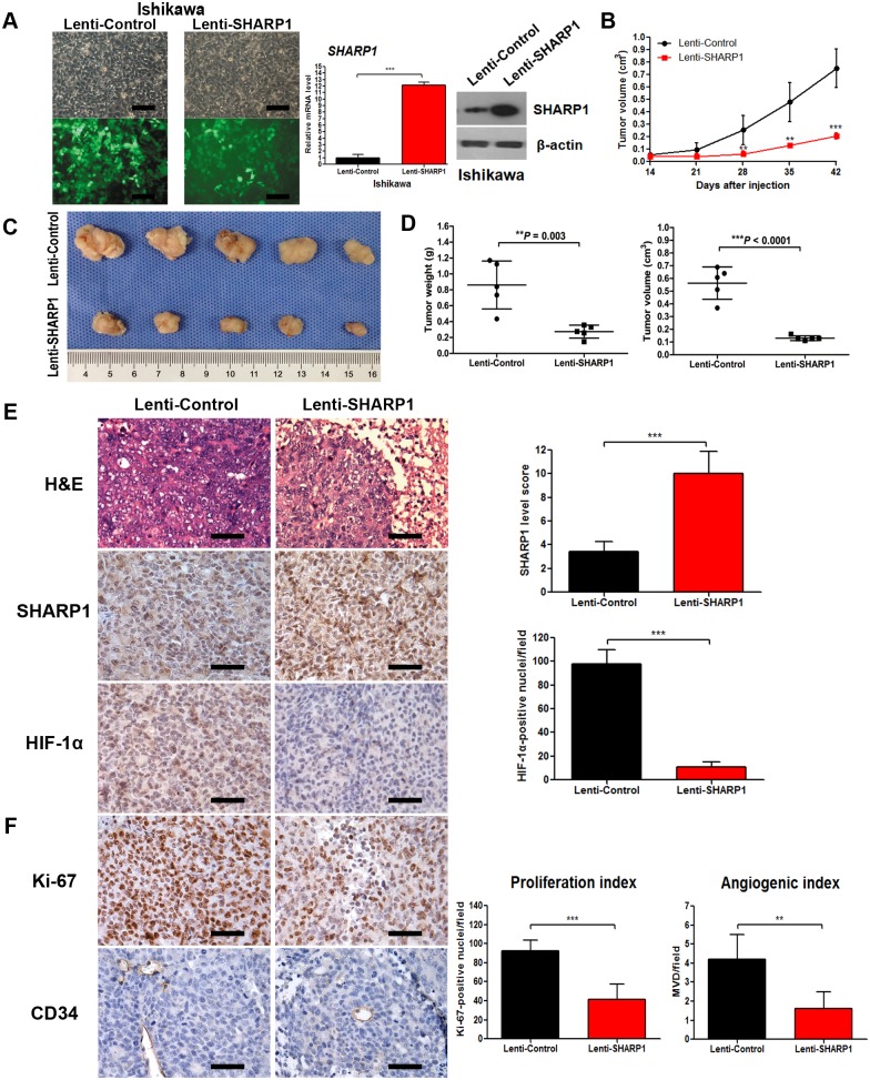
Figure 5. SHARP1 inhibits tumor growth, HIF-1α expression and angiogenesis in tumor xenografts.
(A) Stable transfection of Ishikawa cells with empty lentiviral vectors or lentiviral vectors carrying human SHARP1 gene. Left panels showed morphology of Ishikawa cells under light microscope (upper) or fluorescence microscope (lower) in the same field (magnification: 200×; scale bar: 100 µm), and right panels showed transfection efficiency confirmed by qPCR and western blotting. Ishikawa cells transfected with Lenti-Control or Lenti-SHARP1 were injected subcutaneously into the flank of each mouse. (B) The mean tumor volume was measured by calipers on the indicated days. (C) Photographs of tumors excised 42 days after inoculation of stably transfected cells into nude mice. (D) Tumor weight (left) and volume (right) of each nude mouse at the end of 42 days. (E) Left: Representative microphotographs of H&E, SHARP1 and HIF-1α staining in nude mice tumor tissues (magnification: 400×; scale bar: 50 µm); Right: Statistical analysis of SHARP1 and HIF-1α staining in nude mice tumor tissues. (F) For evaluation of the proliferation index and angiogenic index, the Ki-67-stained nuclei and CD34-positive blood vessels in the hotspot areas were counted at 400× magnification. Representative photographs were taken at 400× magnification (scale bar, 50 µm). Data represent the mean ± SD of 5 grafts in each condition (**P<0.01, ***P<0.001).

