
Figure 5. Histology of week 4 pellet culture in control and chondrogenic media.
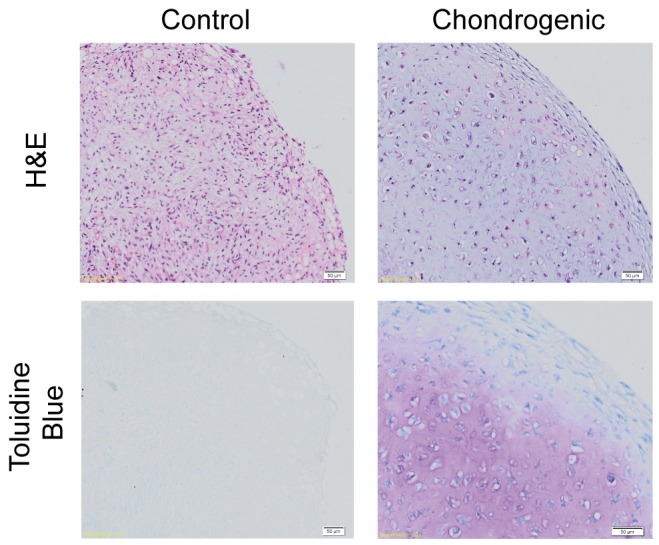
Control media shows cells that appear more fibroblastic in morphology as compared to chondrocytic morphology with extracellular matrix in the chondrogenic pellet. Chondrogenic pellets are larger, well rounded and stains positively for toluidine blue. Magnification 20×. Scale bars at 50 µm as indicated.