
Figure 6. Histology of week 4 chitosan-ASC constructs in chondrogenic and control media.
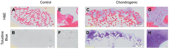
There is a paucity of cellular attachment and growth in control media and lack of toluidine blue staining. Chitosan fibres are clearly stained by eosin. The cells that have grown and attached to the chitosan structure under chondrogenic media shows extracellular matrix deposition with strong toluidine blue staining, suggesting the presence of proteoglycans. Magnification 20×. A–D represent whole tissue images with scale bars at 500 µm as indicated. E–F represents magnified images with scale bars at 20 µm as indicated.