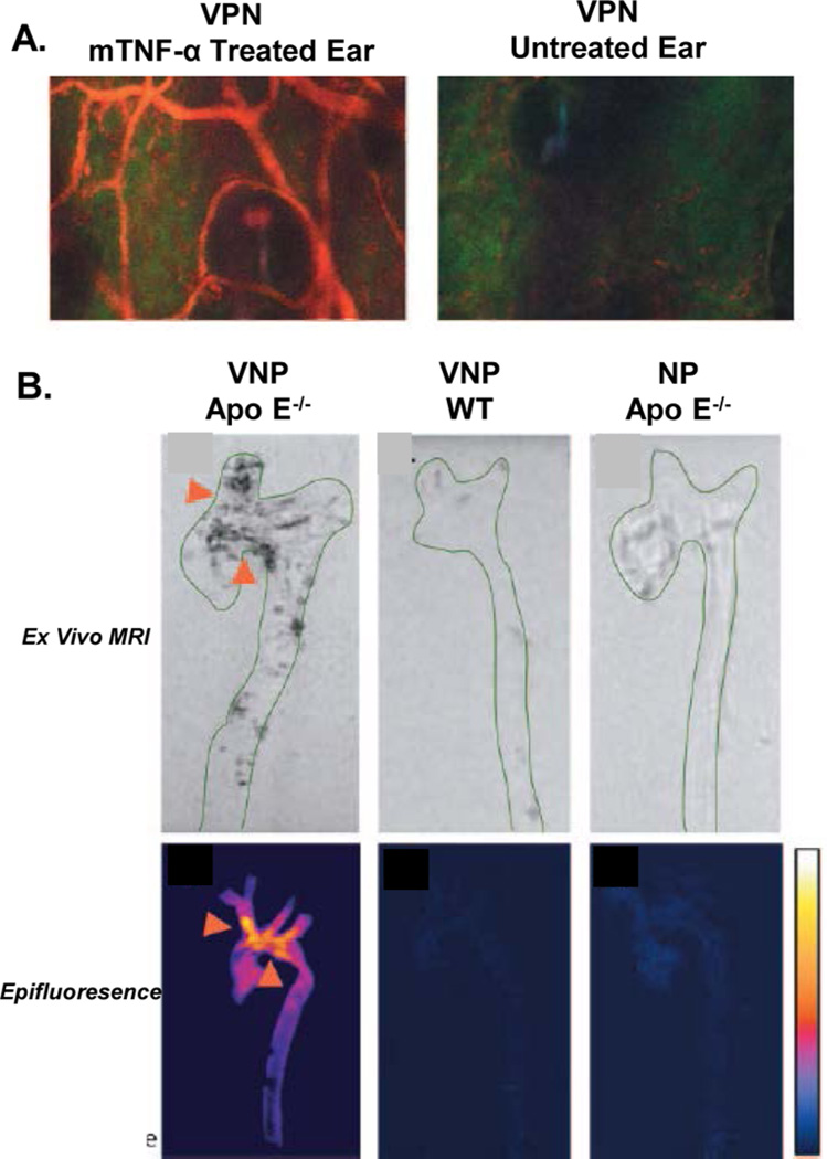
Figure 12.
VCAM-1-specific peptide homes to areas of inflammation and atherosclerotic deposits. (A) The VCAM-1-binding peptide CVHSPNKKCGGSKGK was coupled to a magnetofluorescent nanoparticle (VPN). TNF-α-induced inflammation was induced in one ear of a mouse, while the other was left untreated. Intravital microscopy shows clear accumulation of VPN (red) in the inflamed ear at 4 h, while no binding is observed in the normal control ear. (B) Ex vivo imaging by MR and macroscopic fluorescence at 24 h post intravenous injection detects binding of VPN in animals with atherosclerotic plaques (Apo E−/− mice). No signal is detected in wild-type animals with no plaque development or when a nontargeted nanoparticle is used. The high accumulation of VPN in the aortic arch is indicated by the arrows. Adapted with permission from ref 365. Copyright 2005 American Heart Association Inc.

