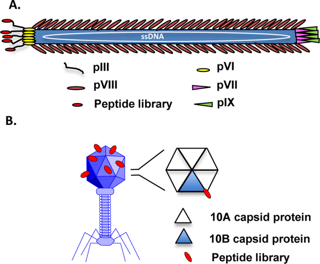
Figure 2.
Filamentous and lytic phage structures. (A) Schematic representation of fd filamentous phage. The random peptide is shown fused to the amino terminus of the pIII coat protein. (B) Representative T7 lytic phage structure. The T7 phage head is comprised of the 10A and 10B capsid proteins arranged as hexamer or pentamer units at a total of 415 proteins per head. A graphical representation of the hexamer capsid unit is shown with a random peptide (red) fused to the 10B protein (blue triangle). T7 phage can be modified to express varying ratios of 10B to 10A protein, displaying peptide sequences in 1–415 copies.

