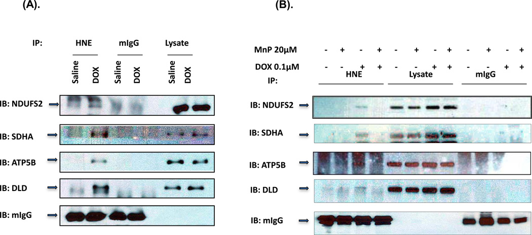
Figure 3. Immunoprecipitation of HNE adducted proteins.
(A) Murine heart homogenates from mice treated with either saline or DOX for 3 days were immunoprecipitated with mouse HNE antibody (lanes 1 & 2) or mouse IgG antibody (lanes 3 & 4) as control. 20 µg of total heart homogenates from IP samples were loaded in lane 5 & 6. The immunoprecipitates were immunoblotted (IB) with antibodies specific for the indicated proteins. The experiment was repeated 3 times to verify results. (B) H9C2 cells pretreated with 20 µM MnP followed with 0.1µM DOX and MnP for 3 days. 500 µg cell lysate was used for each immunoprecipitation. Lane 1–4 were immunoprecipitions with mouse HNE antibody, lane 5–8 were 20 µg of each cell lysate, and lane 9–12 were immunoprecipitations with mouse IgG antibody.

