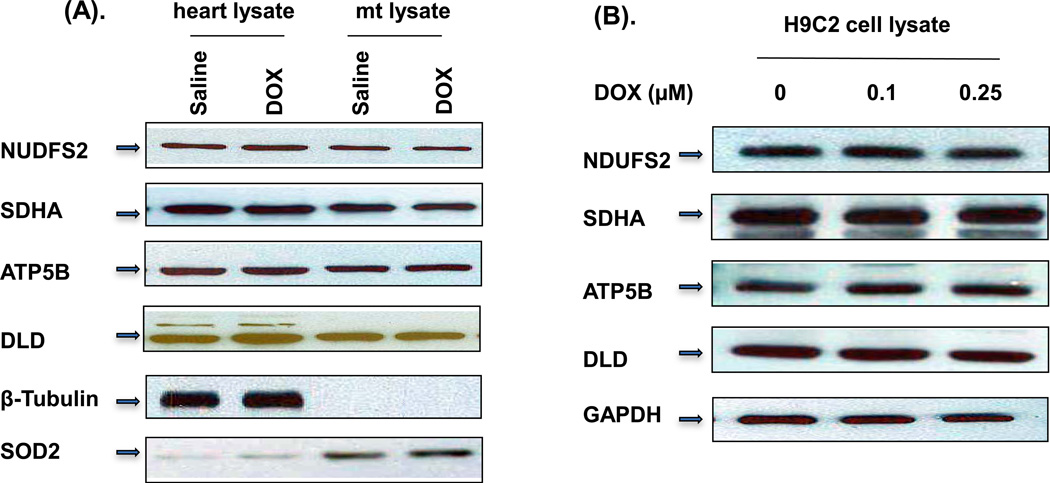
Figure 4. Western blot analysis of the identified HNE adducted proteins.
(A) 30 µg of total mouse heart homogenate and 10 µg of mouse mitochondrial homogenate from 3-days DOX-treated mice were loaded in gels and blotted with different antibodies as indicated. β-Tubulin and SOD2 were used as total heart homogenate and mitochondrial fraction loading controls, respectively. (B) Total H9C2 cell extracts treated with indicated concentration of DOX for 3 days were loaded and blotted with indicated antibodies. The experiments were repeated 4 times.

