
Figure 1.
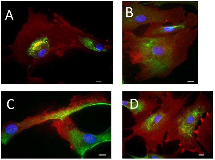
Characterization of VICs. Using explanted human hearts, VICs were isolated from the aortic (A), pulmonary (B), mitral (C), and tricuspid (D) valves. Cells are stained for alphasmooth muscle actin (red), vimentin (green), and nuclear counterstain (blue). All four VICs types exhibit myofibroblast phenotype. Bar = 30μm